Parasites are animals that live on or in another at its expense, steal nutrients, hold down weight gain, and may cause chronic diarrhea, blood in the stool, anemia, skin diseases, and heart disease as well as spreading bacterial, viral, or protozoan diseases. Most have life cycles that carry them through a series of hosts. The object of treatment is not only to destroy the adult parasite but, whenever possible, to break the life cycle .
The parasites of domestic animals fall into two clear-cut groups, internal (endo-parasites) and external (ecto-parasites). Those most frequently found in or on dogs are:
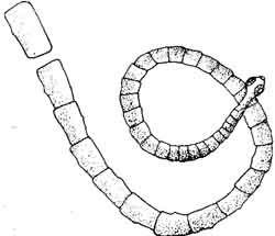
TAPEWORMS
Tapeworms are fairly common but rarely cause any significant problem. Infections can occur when the dog eats uncooked meat, raw freshwater fish, or swallows fleas or lice, all of which may be intermediary hosts. Diagnosis is sometimes made by examining feces microscopically for eggs, but is more frequently the result of finding wriggling segments of the mature worm on top of stools — they look like cut pieces of white ribbon. These can be squashed, releasing packets of eggs for microscopic identification. After an overnight fast, the dog should be given an appropriate medication. Medication for worming is available in pet stores, but it is general — it does not distinguish the different kinds of worms — so a veterinarian’s prescription is much better. Animals can be protected from infection by controlling fleas and lice, and cooking all meat and fish products fed them.
ASCARIDS
Ascarids are probably the most common internal parasites found in young puppies. A pregnant dog can be treated for ascarids; if infected, she should be. Piperazine is the generic name of the medicine. It is not toxic to the fetus — and infections can be transmitted pre-natally, by way of the placenta, or post-natally in the mother’s milk during the first twenty-four hours after birth. During its first month, the pup may also pick up the parasite by eating infected eggs. Lodged in the small intestine, the adult worms compete with the puppy for nutrition, releasing eggs that pass in the feces. In one to four weeks, these contain larval worms that, if the eggs are eaten, can infect a new host such as a rat. The rat too, if eaten, serves as a means of spreading the infection further.

When the larvae are released from the eggs, they spend from two to four weeks traveling through the host body tissues — the lungs and intestinal linings — until, at eight weeks, they reach maturity.
As a result of this migration through the lung tissues, puppies occasionally contract ascarid pneumonia. Emergencies sometimes arise when the worms have become so numerous that they form obstructions in the dog’s system. While these are rare events, severely infected puppies nonetheless develop potbellies and chronic diarrhea, and appear dull.
Diagnosis can result either from spotting the long, spaghetti-like worm in the puppy’s stool or vomit, or by identifying the eggs under a microscope. Three or four treatments with prescribed medication, administered every two to three weeks, will destroy the adult worms, but the larvae are unaffected and must be treated later, when they have developed into adults.
Rarely, children will contract visceral larval migrans by putting their hands in their mouths after having played in the area of dog excrement. The ascarid larvae responsible, toxocara, do not come from dogs directly, how ever, but from the contaminated soil. Without displaying any symptoms directly connected with the worms themselves, infected children may develop pneumonia, liver disease, and even blindness in severe cases. Pets and sandboxes obviously do not go together.
HOOKWORMS
Hookworm infections in young puppies are quite similar to those due to ascarids. The adult worm, in the small bowel, passes its eggs out in the stool. From one to four weeks later, the larvae can gain entry to the host either when the eggs are eaten or by penetrating the skin. Prenatal and postnatal infections occur in the same manner as with the ascarids. Hookworm can be detected in the pregnant mother, but she should not be treated until after the puppies are born, since the treatment would be toxic to the fetus. Prenatal infection can result in blood loss anemias before birth, yet diagnosis through examining the stool is impossible until the dog is approximately eleven days old. The larvae migrate through the lungs before reaching maturity in the small bowel, occasionally causing pneumonia. In addition to competing for nutrition, hookworms feed directly on the dog’s blood, invading the lining of the small bowel. The irritation and damage may result in a severe anemia, blackish diarrhea due to blood loss, and occasionally shock.
Hookworms are rarely passed in the stools, and most diagnoses are the result of microscopic examination of the eggs. Even without this evidence, progressive weakness in a puppy, pallor, and the characteristic diarrhea should cause suspicion, and treatment should be started immediately. Various products your veterinarian can prescribe are effective against the adult hookworm. In cases of severe anemia, blood transfusions, vitamins and other blood-building agents may be used.
WHIPWORMS
Whipworms (Trichuris vulpis), living in the cecum (appendix) and lower bowel, may cause chronic diarrhea containing red blood. The diagnosis can be confirmed by examining the eggs in the stool. Daily administration of prescribed medication for five days will destroy the adult whipworms, but it must be repeated after three months, when the new generation of larvae have reached adulthood.
PROTOZOANS
Protozoans most commonly found in dogs, Giardi canis and coccidia, infect the small bowel. These one-celled organisms cause a watery, mucoid diarrhea, which is plentiful. Identification and treatment must be by a veterinarian. Microscopic identification is necessary; then Kaopectate and bland diets may help in controlling the diarrhea. Giardia can be treated orally with pre scribed medicines twice a day for three days, repeated after three more; antibiotics are used to treat coccidia, administered over a period of twenty-one days.
HEARTWORMS
Heartworm (Dirofilaria imitis) infection, once thought to be confined to the South, has now been identified in virtually every state. Immature microfilana are taken with the blood of infected dogs by various species of mos quito. The mosquito then acts as a carrier, infecting other animals. After migrating within the body, the immature parasites reach maturity in the right side of the heart. Adult worms may range from 8 to 12 inches and a heavy infection can significantly slow circulations through the major blood vessels of the heart and lungs.
Symptoms include coughing, shortness of breath, loss of appetite and weight, and collapse. Diagnosis relies on microscopic identification of the immature parasites in a blood sample. When the disease has been identified, it is treated in the following manner:
(1) Adult heartworms are killed by four daily injections of medication for two successive days while the patient is hospitalized. The dog is then re leased but must rest and be confined, to prevent running, for six weeks. During this period the adult worms die and are trapped by the lungs, where they decompose.
(2) Immature microfilaria are killed when, at the end of the six-week confinement, the dog is started on oral medication. The circulating microfilaria cannot mature unless they develop within a mosquito, but they remain a source of infection. After five to seven days of treatment, the dog’s blood is checked for microfilaria. If they are still present, medication is resumed at a higher dosage.
(3) Re-infection is prevented, once the blood is found free of microfilaria, by giving the dog either liquid medication or pills daily. This will destroy any larvae transmitted by mosquitoes. If you live in an area where heartworms are endemic, your dog should be checked for microfilaria every six months and placed on preventive medication throughout the year. If there are definite seasonal changes, checks should be made at the end of the winter or in the early spring. The animal should receive appropriate medication from spring to early fall.
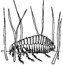
FLEAS
Fleas not only cause dermatitis with their bites, but carry elaborate allergenic toxins in their saliva that increase the reaction to subsequent bites. In severely infected puppies, fleas can cause blood-loss anemia. They also transmit other parasites, such as tapeworms.
The flea remains on the host animal only long enough to feed and mate, spending the rest of its life cycle — from one to six months, though they may live as long as a year — in household rugs and furniture, where its eggs are laid. Fleas are diagnosed by finding either the parasites themselves on your pet, or the black peppery specks called flea dirt lodged among its hairs. Treatment is designed to, first, rid the dog of fleas and, second, prevent re-infestation.
Various products are available to kill the adult fleas, including collars and medallions containing organophosphates; sprays such as Flyte, Norsect spray, and Para-S-Spray; powders; and shampoos. When using these products, watch carefully to see if the animal is sensitive to them, developing skin irritations. Frequent vacuuming, sprays, and professional exterminators can be used as means of ridding the home of fleas.
TICKS
Ticks infesting dogs are usually of two types. The wood tick requires an intermediary host in its life cycle, while the brown tick can spend its entire life on the dog. Ticks spread diseases such as babesiasis, a parasite blood disorder; Rocky Mountain spotted fever; and tick paralysis.
Hardy parasites, ticks are capable of going months without a meal. The female can lay as many as 4,000 to 6,000 eggs at a time. As larvae, these feed on smaller animals, then drop off. After the nymph stage has developed in the soil, it finds a new host. It feeds for from five to seven days, drops off, and Matures, finding yet another host.
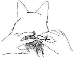
Diagnosis is simply a matter of finding a tick on the dog, usually in the area around the head, neck, and ears. It should be removed by gently pulling it away from the animal. If the head breaks off and remains embedded in the animal, try to remove it with tweezers. If this fails, place hot compresses on the site two or three times daily for a few days. A slight abscess will develop and the body will reject the foreign object. Do not use any caustic sub stances, such as kerosene or lighter fluid, or use burning matches — these can irritate the dog’s skin. To prevent your dog’s getting ticks, use one of the various tick shampoos during the summer months, as directed. If your house has become infested, an exterminator may be essential.
EAR MITES
Ear mites, a common parasite in young dogs, inflame and irritate the tissues of the ear canal. They feed upon the secretions caused by their own irritations. Dogs infected with ear mites will shake their heads and scratch at their ears frequently. Examination of the ears will usually reveal a black, waxy discharge. The tiny white mites can be extracted with a Q-tip and, if there is still doubt, examined under a microscope. A number of mite-killing preparations, if administered every other day for three weeks, will break the mite’s life cycle.
MANGE

SARCOPTIC MANGE. This is caused by a microscopic parasite that infects humans as well as dogs. The mites burrow into the skin, causing irritation, and feed off the secretions that result. This causes intense itching. Young animals are most frequently infected, crusts forming around the ears, eyes, face, and limbs. The heavy chafing and oozing serum often lead to the development of a secondary bacterial pyoderma.
Mites lay from ten to twenty-five eggs, which hatch in three to four days and emerge on the skin surface as six-legged larvae. When they mature, in about fourteen days, the life cycle begins again. Mange mites, larvae, or eggs found on a skin scraping from the affected areas are used for diagnosis.
Applications of products such as lime sulfur and lindane — four to five treatments at intervals of from seven to ten days — are usually effective. These should be under the direction of your veterinarian. Antibiotics and corticosteroids are used to combat secondary bacterial infections and relieve itching until the mites die.
DEMODECTIC MANGE. This is caused by a microscopic parasite that invades the hair follicles of almost all dogs while they are young. Young puppies may acquire demodex through contact with their mothers while they are still nursing. In its milder form, there are small, localized lesions around the face, ears, and legs. But there is no itching. A more severe generalized form is often accompanied by secondary bacterial infections and intense itching.
Skin scrapings from affected areas, containing the mites and their larvae, are the basis for diagnosis. Prescribed medication should be applied daily until the lesions disappear. Dogs with the more resistant generalized form should be treated in the same manner as those with deep skin pyoderma, and given four to five weekly treatments with appropriate medication.
FLIES
Flies, biting the tips of dogs’ ears, cause painful, crusty swellings and infect open wounds with their larvae (maggots). Vaseline applied to the ear tips helps to control the biting. Maggots can be removed by clipping away the hair and cleaning the wound with either hydrogen peroxide or Phisohex. Antibiotic ointments should then be applied to prevent secondary infections.